
Fayil:Schistosoma haematobium egg 4843 lores.jpg

Babu wata babbar saƙa.
Schistosoma_haematobium_egg_4843_lores.jpg (pikisal 700 × 460, girman fayil: 38 KB, irin MIME: image/jpeg)
| Bayani |
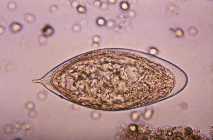
ID#:4843 This micrograph depicts an egg from a Schistosoma haematobium trematode parasite; magnified 500x. Note the egg's posteriorly-protruding, terminal spine, unlike the spinal remnant, which protrudes from the lateral wall of the Schistosoma japonicum egg. These eggs are eliminated in an infected human's feces or urine, and under optimal conditions in a watery environment, the eggs hatch and release "miracidia", which then penetrate a specific snail intermediate host. Once inside the host, the S. haematobium parasite passes through two developmental generations of sporocysts, and are released by the snail into its environment as "cercariae". |
|||
| Rana | ||||
| Masomi | http://phil.cdc.gov/PHIL_Images/20031013/b47fc1793d7443d7a5cdbfbc73d95e53/4843_lores.jpg | |||
| Marubucin | CDC, Public Health Image Library (PHIL) | |||
| Izini (Sake amfani da wannan fayil) |
|
Tarihin fayil
Ku latsa rana/lokaci ku ga fayil yadda yake a wannan lokaci
| Rana/Lokaci | Wadar sufa | Kusurwowi | Ma'aikaci | Bahasi | |
|---|---|---|---|---|---|
| na yanzu | 17:13, 7 Mayu 2006 | | 700 × 460 (38 KB) | Patho | {{Information| |Description=ID#: 4843 Description: This micrograph depicts an egg from the trematode parasite Schistosoma japonicum with its vestigial spine. The Schistosoma japonicum egg is typically oval or subspherical, has a vestigial spine, and is |
Amfani da fayil
Wadannan shafi na amfani wannan fayil:
Amfanin fayil a ko'ina
Wadannan sauran wikis suna amfani da fayil din anan
- Amfani a kan ar.wikipedia.org
- Amfani a kan cs.wikipedia.org
- Amfani a kan de.wikibooks.org
- Amfani a kan en.wikipedia.org
- Amfani a kan fr.wikipedia.org
- Amfani a kan gl.wikipedia.org
- Amfani a kan nl.wikipedia.org
- Amfani a kan sw.wikipedia.org
- Amfani a kan tr.wikipedia.org
- Amfani a kan zh.wikipedia.org